Terms for research-use purchases.
Last updated: September 10, 2025
By accessing or using this website (the “Site”) and purchasing our products, you agree to these Terms. If you do not agree, please do not use the Site.
1) Overview
Welcome to Certified-Pep (“we,” “us,” “our”). By accessing or using this website (the “Site”) and purchasing our products, you agree to these Terms & Conditions (“Terms”). If you do not agree, please do not use the Site.
2) Eligibility & Accounts
You represent that you are 21+ and legally able to enter binding agreements.
You agree to provide accurate information and keep your account secure. We may refuse or cancel orders, or suspend accounts for suspected fraud, misuse, or non-compliance.
3) Research-Use Only (Critical)
All products are for research and laboratory use only, unless explicitly stated otherwise in writing.
Products are not for human or animal consumption, drug use, diagnostic use, clinical use, or incorporation into food, cosmetics, or medical devices.
You are responsible for appropriate handling, storage, and disposal; following MSDS/COA guidance; and complying with all applicable laws, safety standards, and institutional policies.
Statements on the Site are not evaluated by the FDA or any regulatory authority.
4) Ordering, Pricing & Availability
Listings may change without notice. We may limit quantities or discontinue products.
We strive for accuracy, but typographical or system errors (price/availability/specs) may occur. We may cancel or correct such orders and will notify you.
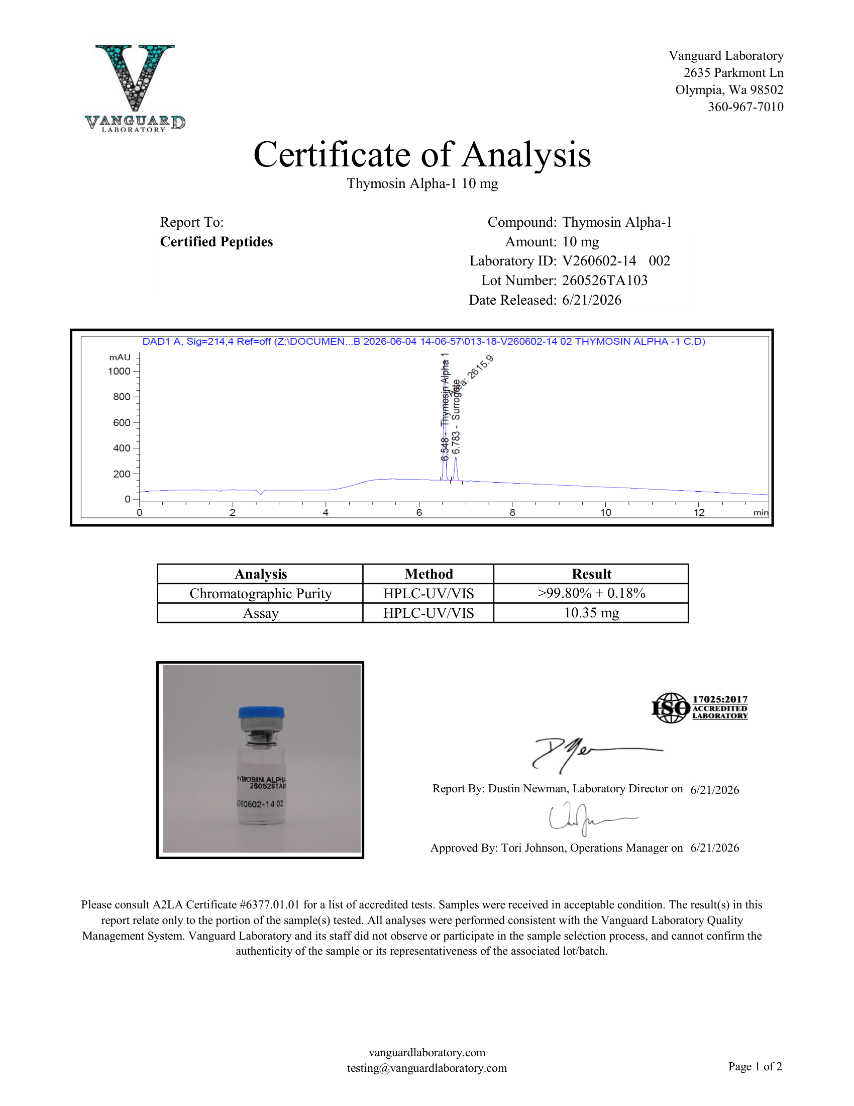
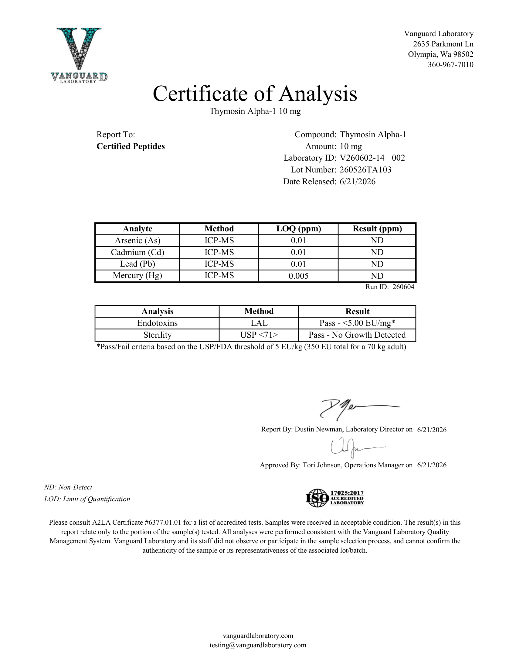
Lot-to-lot variations within normal analytical tolerance may occur. COAs/contaminant reports are provided per lot and reflect measured values for that lot.
5) Payments, Taxes & Chargebacks
Payments are processed by trusted third-party processors. We do not store full card numbers.
Prices are shown in USD unless stated otherwise. Applicable taxes, duties, and fees are your responsibility unless we collect them at checkout.
Suspected payment fraud or misuse of chargebacks may result in account suspension and investigation. We contest improper chargebacks.
6) Shipping & Risk of Loss
We aim to ship promptly. Delivery times are estimates and may vary by carrier, weather, holidays, or customs.
Risk of loss transfers to you when the carrier accepts the shipment. Consider purchasing route/carrier insurance where available.
Ensure your shipping address is accurate. We are not responsible for delays or losses due to incorrect addresses or carrier issues.
International: You are responsible for import permits, duties/taxes, and compliance with local regulations. We are not liable for customs delays, holds, seizures, or destruction.
7) Inspection & Acceptance
Inspect shipments upon delivery. Report shipping damage, shortages, or incorrect items within 3 business days of receipt with photos and order details.
After that window, the order is deemed accepted.
8) Returns & Refunds
All sales are final. Due to the nature of research products, we cannot accept returns or provide refunds after purchase.
9) Limited Warranty & Disclaimers
We warrant that, at the time of shipment, products will conform to our specifications for the applicable lot.
Your exclusive remedy for breach of this warranty is, at our option, replacement of the product or refund of the purchase price for the affected item.
Disclaimers: Except for the foregoing, products are provided “AS IS” and “AS AVAILABLE,” without warranties of merchantability, fitness for a particular purpose, or non-infringement.
10) Limitation of Liability & Indemnity
To the maximum extent permitted by law, we are not liable for indirect, incidental, special, consequential, or punitive damages (including lost profits, data, or use), even if advised of the possibility.
Our total liability for any claim related to a purchase will not exceed the amount you paid for the product giving rise to the claim.
You agree to indemnify and hold us harmless from claims arising from your handling, storage, use, or resale of products, or your breach of these Terms.
11) Bundles, Quantity & Promotions
Bundle prices apply only when purchasing the specific bundle SKU. Adding multiple units of a single product does not grant the bundle price unless the bundle product is added to cart.
Coupons/discounts cannot be combined unless stated. We may modify or end promotions at any time.
12) Site Use, Content & Intellectual Property
All Site content (text, graphics, logos, product images, COAs) is owned by or licensed to us and protected by law.
You may not copy, scrape, frame, reverse-engineer, or use automated tools to extract content without our written consent.
User reviews or submissions must be lawful and non-infringing; by submitting, you grant us a non-exclusive license to display and use them on the Site.
13) Compliance, Resale & Export
You are responsible for compliance with all applicable laws and institutional rules regarding purchase, possession, use, resale, transport, and disposal.
You will not export or re-export products in violation of export control or sanctions laws.
14) Governing Law & Disputes
These Terms are governed by applicable federal and state laws of the United States, without regard to conflict-of-law principles.
Any dispute arising out of or relating to these Terms or use of the Site shall be handled by a court of competent jurisdiction, except where otherwise required by applicable law. Small-claims actions may be brought where permitted by law.
15) Changes, Severability & Assignment
We may update these Terms at any time by posting the revised version on this page with a new “Last updated” date.
If any provision is found unenforceable, the remainder will remain in effect.
We may assign our rights/obligations; you may not assign without our consent.
16) Contact
Questions about these Terms?
Email: [email protected]
For faster assistance, you may also contact our support team through Live Chat on the Certified-Pep website.
By completing a purchase, you acknowledge that you have read, understood, and agree to these Terms & Conditions.